
Radiofrequency ablation of intra-articular osteoid osteoma of the hip.
J Int Med Res. 2006 Sep-Oct;34(5):537-44.
Papagelopoulos PJ, Mavrogenis AF, Kyriakopoulos CK, Benetos IS, Kelekis NL, Andreou J, Soucacos PN.
Diagnosis and treatment of intra-articular osteoid osteoma is challenging. We present 16 patients with intra-articular osteoid osteomas of the hip treated with percutaneous radiofrequency ablation. Eight osteoid osteomas were located in the femoral head, six in the femoral neck, and two in the acetabulum. Three of the 16 patients had had an incorrect previous diagnosis. Percutaneous radiofrequency ablation was a clinical and technical success in all 16 patients. Within the first 24 h after the procedure, pain improved in all patients. Five patients had pain relief within the first 3 days after the procedure, nine patients within the first week and two patients within 2 weeks. Residual or recurrent symptoms were not reported by the last follow-up. At the 12-month follow-up, computed tomography and magnetic resonance imaging showed complete ossification and bone regeneration at the site of the lesion in three patients, partial ossification in six patients and no changes in seven patients. Computed tomography-guided percutaneous radiofrequency ablation is a simple, minimally invasive, safe and effective method for the treatment of most intra-articular osteoid osteomas.
News
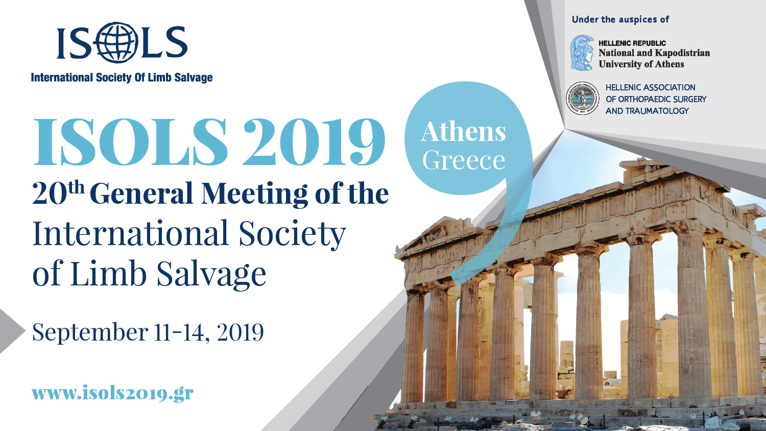 ISOLS 2019 20th General Meeting of the International Society of Limb Salvage Athens, Greece September 11th -14th, 2019 |
Useful Links
|
• American Academy of Orthopaedic Surgeons, www.aaos.org • Mayo Clinic, www.mayoclinic.org • Connective Tissue Oncology Society, www.ctos.org • European MusculoSkeletal Oncology Society (EMSOS), www.emsos.org • MusculoSkeletal Tumor Society (MSTS), www.msts.org • International Society of Limb Salvage (ISOLS), www.isols.org |
Contact Information
|
Athens University Musculoskeletal Tumor Center Sec: Heleni Ziavrou Email: elziavrou@gmail.com Tel: +30.210.5832399, +30.210.5832398 Fax: +30.210.5832735 Athens University General Hospital "ATTIKON" 1, Rimini Str, 124 62, Haidari Athens, Greece |